
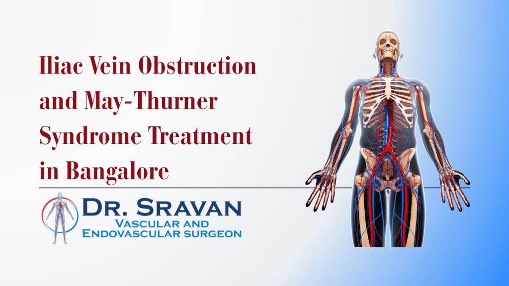
Persistent swelling of one leg is sometimes caused by a problem higher in the venous system, where the large iliac veins carry blood from the legs through the pelvis. May-Thurner syndrome is one form of iliac vein obstruction in which the left iliac vein is compressed by the neighbouring right iliac artery and the spine.
However, seeing iliac-vein compression on a scan does not automatically mean that it needs treatment. Some people have anatomical compression without symptoms. I look for a clear connection between the vein obstruction and problems such as significant one-sided swelling, previous extensive DVT, heaviness, venous skin changes or ulceration before considering treatment such as iliac vein stenting.
What Is May-Thurner Syndrome?
May-Thurner syndrome is a condition in which a major vein in the pelvis is compressed.
In the classic pattern, the left common iliac vein passes between the right common iliac artery and the spine.
The artery can press against the vein and reduce the space available for blood to return from the left leg.
This can create:
- slower venous drainage
- increased pressure in the leg veins
- chronic swelling
- heaviness or discomfort
- development of collateral veins
- increased susceptibility to deep vein thrombosis in some patients
The Society for Vascular Surgery describes the condition as compression of the left iliac vein that can produce chronic leg symptoms or, in more severe cases, DVT.
But the anatomy needs to be interpreted carefully.
Is Iliac Vein Compression Always May-Thurner Syndrome?
Not necessarily.
Some degree of iliac-vein compression can be seen in people who have no symptoms.
Finding narrowing on CT or another scan does not prove that the narrowing is responsible for a patient’s leg problem.
That distinction matters because an incidental anatomical finding should not automatically lead to a procedure.
I prefer to separate three questions:
- Is an iliac vein narrowed or obstructed?
- Is the patient having symptoms that fit this problem?
- Is the obstruction significant enough that treating it is likely to provide meaningful benefit?
Only after answering all three does venous stenting become a reasonable discussion.
Current venous guidance similarly supports stenting only in selected patients with symptomatic iliocaval obstruction rather than treating every anatomical narrowing.
Is May-Thurner Syndrome the Only Cause of Iliac Vein Obstruction?
No.
May-Thurner syndrome is one important cause, but iliac-vein obstruction is a broader problem.
Two major patterns are commonly considered.
Non-thrombotic iliac vein compression
This includes May-Thurner-type anatomical compression.
The vein is narrowed from outside rather than being permanently damaged by a previous clot.
Post-thrombotic iliac vein obstruction
A previous DVT can damage the vein.
After the acute clot settles, the vein may remain:
- narrowed
- scarred
- irregular
- partially blocked
- completely chronically occluded
This can interfere with blood returning from the leg and contribute to post-thrombotic syndrome.
Some patients have a combination of previous thrombosis and anatomical compression.
That is why the history of an earlier DVT is particularly important.
What Symptoms Can Iliac Vein Obstruction Cause?
Symptoms depend on how severe the obstruction is and how effectively the body has developed alternative venous pathways.
Possible symptoms include:
- persistent swelling of one leg
- heaviness
- aching
- tightness
- a feeling of pressure in the leg
- discomfort after prolonged standing
- symptoms that worsen as the day progresses
- prominent superficial veins
- venous skin changes
- recurrent venous ulceration in advanced disease
Some patients describe venous claudication.
This is a bursting, tight or heavy discomfort that develops during walking or exercise because venous blood cannot leave the leg efficiently.
It is different from arterial claudication, which is caused by inadequate blood entering the leg through the arteries.
Why Does May-Thurner Syndrome Commonly Affect the Left Leg?
The classic anatomy involves the right common iliac artery crossing over the left common iliac vein.
The vein is compressed between the artery and the spine.
For this reason, unexplained or recurrent swelling of the left leg can raise suspicion for iliac vein compression.
However, other forms of iliac-vein obstruction can affect the right side or both sides.
A diagnosis should therefore not be made simply because swelling is on the left.
Can May-Thurner Syndrome Cause DVT?
Yes.
Significant compression can slow venous blood flow and may contribute to development of a clot in the deep veins.
Some patients are first diagnosed with May-Thurner syndrome after developing an extensive DVT involving the thigh or pelvic veins.
This is called iliofemoral DVT when the clot involves major proximal veins.
If you have new swelling, pain or warmth in one leg, DVT needs to be considered before assuming that symptoms are simply due to chronic vein compression.
Read the DVT treatment guide explaining how clot location and severity affect treatment.
Can May-Thurner Syndrome Occur Without DVT?
Yes.
A patient can have clinically significant iliac-vein compression without having an acute blood clot.
Symptoms may develop gradually and include:
- chronic swelling
- heaviness
- aching
- venous skin changes
- visible collateral veins
However, this is also the group in whom careful patient selection becomes particularly important.
Because anatomical compression can exist without causing problems, treatment should be based on symptoms plus clinically meaningful obstruction, not imaging alone.
Why Is My Leg Still Swollen After a DVT?
Persistent swelling after DVT does not automatically mean that another clot has formed.
Several mechanisms can contribute.
Residual obstruction
The vein may remain narrowed or partially blocked after the acute clot.
Scar tissue inside the vein
Healing after thrombosis can leave fibrotic material inside the vessel.
Valve damage
A DVT can permanently damage the valves that normally prevent blood from flowing backwards.
Iliac vein obstruction
A major pelvic vein may remain chronically narrowed or obstructed.
Combined obstruction and reflux
Some patients have both poor venous outflow and damaged venous valves.
These chronic changes can cause post-thrombotic syndrome with swelling, heaviness, discomfort, skin changes and sometimes venous ulcers.
For broader information about chronic problems after DVT, see the Deep Vein Disorders page.
When Should Persistent Leg Swelling Be Investigated Further?
Many conditions can cause leg swelling.
These include:
- superficial venous insufficiency
- previous DVT
- lymphedema
- infection
- medication effects
- heart, kidney or liver disease
- injury
- reduced mobility
Iliac-vein obstruction becomes more relevant when there is:
- marked swelling mainly affecting one leg
- persistent swelling after an iliofemoral DVT
- swelling involving the thigh as well as the lower leg
- significant heaviness or venous claudication
- recurrent DVT affecting the same leg
- unexplained chronic venous skin changes
- venous ulceration despite appropriate treatment
- imaging suggesting pelvic-vein obstruction
The pattern of symptoms should guide the investigation.
How Is May-Thurner Syndrome Diagnosed?
There is no single test that answers every question.
The evaluation usually progresses from clinical assessment to imaging of the deep venous system.
Clinical Assessment
I first want to understand:
- which leg is swollen
- when the swelling started
- whether it is constant or varies during the day
- whether there has been a previous DVT
- where that DVT was located
- whether symptoms improved after anticoagulation
- whether there is heaviness or pain with walking
- whether varicose veins or skin changes have developed
- whether there has been a venous ulcer
- whether previous venous procedures have been performed
Both legs are examined for differences in swelling, skin appearance and superficial venous patterns.
Venous Doppler Ultrasound
Ultrasound is usually an important first investigation for leg-vein symptoms.
It can identify:
- acute DVT
- previous thrombotic changes
- venous reflux
- abnormalities in accessible deep veins
However, the iliac veins lie deep inside the pelvis.
Routine leg Doppler ultrasound may therefore not fully assess every pelvic-vein obstruction.
A normal leg Doppler does not automatically exclude May-Thurner syndrome when the symptoms and history strongly suggest a higher venous obstruction.
Read more about what different venous scans can assess in the Vascular Doppler Scan guide.
CT Venography
CT venography can provide a wider view of the pelvic veins.
It may help show:
- compression of the iliac vein
- chronic obstruction
- collateral veins
- relationship between arteries and veins
- other possible causes of pelvic venous narrowing
The decision to use contrast imaging also takes kidney function and other medical factors into account.
MR Venography
MR venography can provide another method of evaluating pelvic venous anatomy in selected patients.
The most appropriate scan depends on the clinical question and the patient’s circumstances.
Catheter Venography
Venography is an invasive test performed through a vein.
Contrast is injected while X-ray images show how blood travels through the pelvic venous system.
It may demonstrate:
- narrowing
- delayed drainage
- chronic occlusion
- collateral pathways
Venography may form part of the treatment procedure when intervention is being considered.
What Is IVUS and Why Is It Used?
IVUS means intravascular ultrasound.
Instead of looking at the vein from outside the body, a very small ultrasound probe is passed through the vein.
This allows the vascular specialist to examine the vessel from inside.
IVUS can help assess:
- the true size of the vein
- degree of narrowing
- length of the abnormal segment
- post-thrombotic scar tissue
- the areas that need to be covered if a stent is placed
Research comparing venography and IVUS has shown that standard venography can underestimate some iliac-vein obstructions. IVUS can therefore provide valuable additional information when planning treatment.
This does not mean every patient with leg swelling needs IVUS.
It is generally considered when there is a strong clinical reason to investigate or treat a significant deep venous obstruction.
Does Every Patient With May-Thurner Syndrome Need Treatment?
No.
A patient with incidental iliac-vein compression and no meaningful symptoms may not require a procedure.
The Society for Vascular Surgery notes that asymptomatic patients generally do not need intervention, while treatment may be considered when symptoms become significant.
The decision becomes more relevant when a confirmed obstruction is associated with problems such as:
- significant chronic swelling
- disabling heaviness or pain
- venous claudication
- severe post-thrombotic symptoms
- venous skin damage
- recurrent venous ulceration
- extensive DVT associated with an underlying obstruction
The key word is symptomatic.
Treat the patient, not the scan.
How Is Iliac Vein Obstruction Treated?
Treatment depends on whether the problem is:
- mild or severe
- acute or chronic
- thrombotic or non-thrombotic
- producing DVT or only chronic symptoms
Several approaches may be used.
Conservative Treatment
For mild symptoms, treatment may include:
- regular movement
- avoiding prolonged immobility
- leg elevation where appropriate
- compression when suitable
- management of other venous disease
- weight management where relevant
- treatment of additional clotting risk factors
Conservative treatment may be reasonable when symptoms are limited and there is no urgent venous complication.
If symptoms remain significant despite appropriate conservative management and clinically meaningful iliac obstruction is confirmed, intervention may then be considered. Current SCAI guidance similarly supports iliocaval stenting in selected symptomatic patients, while recognising that the evidence base is not equally strong for every patient group.
Treatment When Acute DVT Is Present
If May-Thurner syndrome is discovered during an acute DVT, the immediate priority is treatment of the thrombosis.
Anticoagulation is the main treatment for most DVT patients.
Selected patients with extensive iliofemoral thrombosis and severe symptoms may be considered for additional catheter-based clot treatment.
These procedures may include:
- mechanical thrombectomy
- catheter-directed thrombolysis
- other thrombectomy techniques
If a clinically important underlying iliac obstruction remains after the acute clot has been treated, venous stenting may then be considered.
Not every DVT requires clot removal or a stent.
The DVT treatment page explains how this decision is made.
What Is Iliac Vein Angioplasty?
Venous angioplasty uses a balloon to widen the obstructed section of vein.
The principle resembles arterial angioplasty, but veins behave differently from arteries.
With significant iliac-vein compression or chronic scar-related obstruction, balloon treatment alone may not provide enough lasting support.
A venous stent may therefore be required when intervention is appropriate.
What Is an Iliac Vein Stent?
A venous stent is a flexible mesh tube placed inside the obstructed iliac vein.
Its purpose is to support the vein and create a more adequate channel for blood returning from the leg.
During treatment:
- Venous access is obtained through an appropriate vein.
- Venography and sometimes IVUS are used to assess the obstruction.
- A guidewire is passed through the narrowed or blocked segment.
- The vein is dilated when appropriate.
- A stent is positioned across the clinically significant obstruction.
- The result is reassessed to confirm appropriate flow through the treated segment.
The procedure is performed using catheter-based endovascular techniques.
Will a Venous Stent Cure Leg Swelling?
Not necessarily.
A stent can improve venous outflow when a significant iliac obstruction is responsible for symptoms.
But leg swelling can have several causes.
Even in someone with a genuine iliac obstruction, there may also be:
- damaged deep-vein valves
- superficial venous reflux
- lymphatic dysfunction
- obesity
- reduced mobility
- chronic tissue changes
This means symptoms may improve without disappearing completely.
That is why I would not recommend stenting based only on the hope that every swollen leg will return completely to normal.
The expected benefit should be discussed realistically before treatment.
What Are the Risks of Iliac Vein Stenting?
Venous stenting is minimally invasive, but it is still an invasive vascular procedure.
Possible risks include:
- bleeding or bruising at the access site
- vein injury
- blood clot formation
- stent thrombosis
- narrowing within or around the stent
- incomplete symptom improvement
- need for another venous procedure
- contrast-related problems
- complications related to anticoagulant or antiplatelet treatment
- uncommon stent-related complications
Long-term follow-up is important because a venous stent remains inside the body.
The expected benefit needs to justify these risks.
Do I Need Blood Thinners After an Iliac Vein Stent?
Many patients require antithrombotic medication after venous stenting, particularly when DVT or post-thrombotic disease is involved.
However, there is no single medication plan that should be copied for every patient.
The choice and duration can depend on:
- whether there was an acute DVT
- whether the obstruction is post-thrombotic
- previous episodes of thrombosis
- bleeding risk
- other medical conditions
- the intervention performed
- the treating vascular team’s protocol
Do not stop or alter anticoagulants or antiplatelet medicines without discussing the change with the treating doctor.
What Happens After Iliac Vein Stenting?
Follow-up may include assessment of:
- leg swelling
- heaviness and pain
- ability to walk
- skin changes
- venous ulcer healing where relevant
- stent flow
- recurrence of symptoms
- medication tolerance
Patients may also receive advice about:
- mobility
- compression
- medication
- activity
- follow-up imaging
New or rapidly increasing swelling after treatment should be reassessed.
Can Iliac Vein Obstruction Cause Varicose Veins?
It can contribute to abnormal venous pressure and development of collateral veins.
However, ordinary superficial venous reflux is a much more common reason for varicose veins.
When significant deep venous obstruction and superficial reflux coexist, treatment planning becomes more complex.
The deeper obstruction needs to be understood before assuming that treating visible surface veins alone will solve the patient’s symptoms.
Can Iliac Vein Obstruction Cause a Venous Ulcer?
Severe chronic venous hypertension can contribute to:
- swelling
- pigmentation
- hardening of the skin
- inflammation
- eventually ulceration
This may occur after extensive DVT or significant chronic outflow obstruction.
However, venous ulcers can also result from superficial venous reflux or combined venous problems.
The cause should be mapped before choosing treatment.
When Is One-Sided Leg Swelling an Emergency?
Persistent chronic swelling can usually be evaluated through a planned vascular consultation.
Sudden one-sided swelling is different.
Seek urgent medical assessment if one leg suddenly becomes:
- significantly swollen
- painful
- unusually warm
- markedly discoloured
These symptoms can occur with acute DVT.
If leg symptoms occur with:
- sudden breathlessness
- chest pain
- coughing blood
- fainting
- severe breathing difficulty
seek emergency medical care immediately because these can occur with pulmonary embolism.
Do not wait for a routine outpatient consultation in that situation.
Iliac Vein Obstruction Treatment With Dr Sravan C.P.S
Dr Sravan C.P.S is a vascular and endovascular surgeon in Bangalore whose work includes deep vein disorders, DVT, chronic venous disease and endovascular vascular treatment.
His verified qualifications include:
- MBBS
- MS in General Surgery
- MRCS from the Royal College of Edinburgh, UK
- DNB in Peripheral Vascular Surgery
- FEVS from National University Hospital, Singapore
He has also served as an Assistant Professor of Vascular Surgery at Sri Jayadeva Institute of Cardiovascular Sciences and Research.
The important decision in suspected May-Thurner syndrome is not:
“The CT says the vein is compressed. Should we put in a stent?”
I would first want to answer:
- Are the symptoms truly coming from venous obstruction?
- Is the obstruction acute, chronic or post-thrombotic?
- Has there been a previous iliofemoral DVT?
- Is superficial reflux also present?
- Is the obstruction clinically significant?
- Have appropriate conservative measures been considered?
- Would improving iliac-vein outflow realistically improve the patient’s symptoms?
- Does the anatomy support safe and useful stenting?
- What follow-up and antithrombotic treatment would be required afterwards?
That prevents an anatomical finding from being treated as though it were automatically a disease requiring intervention.
Read more about Dr Sravan’s vascular and endovascular training.
When Should You See a Vascular Surgeon?
Consider vascular evaluation if you have:
- persistent swelling mainly affecting one leg
- significant left-leg swelling without a clear explanation
- a previous extensive DVT involving the thigh or pelvic veins
- swelling that continues long after DVT treatment
- recurrent DVT in the same leg
- heaviness or pain that worsens with prolonged standing
- venous claudication
- chronic skin changes after DVT
- a recurrent venous ulcer
- CT or MRI reporting significant iliac-vein compression
- another doctor suggesting iliac-vein angioplasty or stenting
- uncertainty about whether a scan finding actually explains your symptoms
The purpose of consultation is to establish whether a clinically important venous obstruction is present before choosing a procedure.
For non-emergency symptoms, book a vascular consultation with Dr Sravan in Bangalore.
Frequently Asked Questions About May-Thurner Syndrome and Iliac Vein Obstruction
Is May-Thurner syndrome the same as having a compressed iliac vein on a scan?
Not always. Some people have anatomical iliac-vein compression without meaningful symptoms. May-Thurner syndrome becomes clinically relevant when the compression is associated with problems such as significant leg swelling, pain, venous changes or DVT. A scan finding alone does not automatically mean treatment is necessary.
Does May-Thurner syndrome always cause DVT?
No. Some patients develop chronic swelling or heaviness without ever having an acute DVT. Others are first diagnosed after an extensive left-leg DVT. The symptoms and treatment depend on the degree of obstruction and whether thrombosis is present.
Can Doppler ultrasound diagnose May-Thurner syndrome?
Doppler ultrasound is useful for detecting DVT and assessing many leg-vein problems, but the iliac veins lie deep in the pelvis and may not always be completely assessed with routine ultrasound. CT venography, MR venography, catheter venography or IVUS may be needed when significant pelvic-vein obstruction is suspected.
Does every patient with May-Thurner syndrome need a stent?
No. Patients without meaningful symptoms may not require intervention. Venous stenting is generally considered for selected patients when a significant iliac obstruction is clearly related to troublesome symptoms, post-thrombotic disease or other clinically important venous complications.
Why is my leg still swollen months after a DVT?
A previous DVT can leave scar tissue, chronic obstruction or damaged venous valves. This can cause post-thrombotic syndrome. In selected patients, persistent iliac-vein obstruction contributes significantly and may warrant further imaging and specialist assessment.
Will an iliac vein stent make my leg swelling completely disappear?
It cannot be guaranteed. Symptoms may improve if significant iliac obstruction is a major cause of the swelling, but other problems such as valve damage, superficial venous reflux or lymphatic dysfunction may remain. Treatment should be recommended only when a meaningful benefit is reasonably expected.
Do I need lifelong blood thinners after iliac vein stenting?
Not every patient follows the same medication plan. The type and duration of antithrombotic treatment depend on factors such as previous DVT, post-thrombotic disease, recurrent clotting risk, bleeding risk and the procedure performed. Medication decisions should be individualised by the treating vascular team.
Conclusion
May-Thurner syndrome is one possible cause of iliac-vein obstruction, but an anatomical compression seen on a scan should not automatically lead to venous stenting.
The clinically important question is whether the obstruction is preventing blood from leaving the leg efficiently enough to cause significant symptoms or complications.
This becomes particularly relevant in patients with persistent one-sided swelling, previous iliofemoral DVT, post-thrombotic symptoms, venous skin damage or recurrent ulceration.
Assessment may include Doppler ultrasound, CT or MR venography, catheter venography and, in selected cases, intravascular ultrasound.
When a significant symptomatic iliac obstruction is confirmed, treatment may range from conservative management to venous angioplasty and stenting. If acute DVT is also present, anticoagulation and selected clot-removal strategies may become part of the treatment pathway.
Opening a narrow vein is not the point on its own.
What matters is identifying which obstruction is clinically important, whether correcting it is likely to help, and which treatment fits that individual patient.
